
 In Search of a Physics of Cytoplasm
John Grant Watterson
18 Tomanbil Terrace, Ashmore, 4214 Queensland. Australia
Email: jgwatterson@gmail.com
(retired: formerly of Faculty of Science and Engineering, Griffith University, Griffith University, Queensland, Australia)
Keywords: cytoplasm, gel, water structure, protein structure, molecular machine
Received: February 2, 2018; Revised: May 24, 2018; Accepted: May 25, 2018; Published: Date July 15, 2018; Available Online: July 15, 2018
Abstract
At the present time we do not have a theoretical understanding of the physics of cytoplasm. For the greater part of the last century, cell biologists reflexively accepted the physicochemistry of the cell as an extension of reductionist classical thermodynamics. This meant that the physics of the cell was viewed as a collection of random events obeying the laws of statistical mechanics, and its chemistry viewed essentially as a collection of test-tube reactions. During the 1990s however, this general view became overshadowed, and eventually replaced, by more holistic approaches. The awarding of the Nobel Prize to Boyer and Walker in 1997 for mapping the molecular structure and function of ATP synthase carried with it the implication that biologists of all persuasions accepted their model, thereby legitimizing molecular modeling of protein machines as a scientific method. However, biologists utilizing these methods ignore basic energetic questions concerning the role of the medium in stabilizing their machines, and even more importantly, the medium’s role as an active participant in molecular mechanisms. On the reverse side of the same proverbial coin, the failure of the IBM Blue Gene Project launched in 1999 to solve protein structure using the classical approach of free energy minimization, revealed the inability of thermodynamics to answer questions concerning the inner workings of biological processes. Here too, the approach ignored water’s role in generating physical forces exerted at the molecular level. In this review, I highlight fundamental problems with the current understanding of cytoplasm requiring urgent attention from physicists willing to approach the topic from a new perspective.
Introduction
Cytoplasm is the living substance, so to have a scientific understanding of life processes, we need to understand how it works. According to deterministic theories, knowledge of any system is gained by studying its parts. We find this view expressed in uncompromising terms by prominent physicists such as Feynman (1990) or Gell-Man (1994) in reference to biology. In this view, the properties of the biological cell can be reduced to the thermodynamics of its component molecules, implying that the physics of cytoplasm is the physics of statistical mechanics, and by extension, the chemistry of cytoplasm is the chemistry of a random collection of molecular collisions in solution. But few biologists today would agree that any common biological activity, say cell division, is a collection of random events. It’s the result of an astronomical number of chemical steps proceeding under spatiotemporal control, which must synchronize with steps exerting mechanical forces in order to direct the necessary local molecular displacements. So in practice, biologists ignore thermodynamic principles when building models of how proteins perform work, as is well illustrated by the host of complex mechanisms we find depicted on the Protein Data Bank poster “Molecular Machinery” (Web ref 1.). And indeed, because of the volume of research published in support of the biologist’s proposals gathered since the 1990s, thermodynamic objections to the mechanical models have been largely ignored over the past two decades. However, in a strident reaction to results of recent observations obtained in the newer fields of bio-sensing where the cell responds to external stimuli, the controversy has now resurfaced in a dramatic way.
Old Physics vs New Observations
In his recent article, the thermodynamicist, Meister (2016), launched a scathing attack on publications by groups reporting observations that nanosized cellular ferromagnets could detect the earth’s magnetic field; i.e., observations of magnetic biosensors (Stanley et al., 2016; Qin et al., 2016; Wheeler et al., 2016). He draws on quantitative comparisons between the physical strengths (energies, forces, torques) that would be expected from results claimed by the groups, and strengths which are quickly and easily calculated from the basic laws of physics. His numerical values show that the claimed effects must be weaker, by many orders of magnitude, than the disruptive influence that thermal motion must have on their magnets according to physical laws. With stern words for the editor and referees of Nature, he asserts the reports should not have been published, because firstly, the several biological explanations proposed by the authors must all be wrong, and secondly, the publications have therefore discouraged younger scientists from doing research in the field in the future.
Meister’s underlying argument that the claims “conflict with the basic laws of physics,” is based on the principle of the equipartition of thermal energy, kT, with respect to the degrees of freedom of molecular orientational motion, which must apply to the molecular constituents of living cells. To use his words again, a molecular magnet must be subject to the “thermal forces that randomize its orientation.” This is in line with the overriding view of the cell as a solution of protein catalysts (enzymes) that carry out its metabolic chemistry. In other words, the view once held by physical scientists generally of the cell as essentially a bag of enzymes, is here still taken as fact.
This picture of the cell is not correct. Today, that the internal medium of the cell is a gel is now well established. In addition, the physicochemistry of the cell is not random, or even reversible. And contrary to the principles of statistical mechanics, it is exclusively unidirectional. Let us recall some of the historical background that has led to the view of the cell accepted by biologists today.
History of the Medium
When the famed microscopist, Frey-Wyssling, examined various cell types in his light microscope, he discovered a puzzle. The cell interior appeared clear, as a solution, but it displayed the properties of a solid (Frey-Wyssling, 1940). In a perceptive description of this observation, he used the term “liquid crystal” in the year 1940! Later he warned against viewing the cell interior as a clear solution in the fluid state because of “the double nature of the cytoplasm; it is solid and liquid at the same time” (Frey-Wyssling, 1948). The later technique of electron microscopy showed some tissues in such regular alignment that they could be described as crystalline; a description validated as early as 1961 by the observation that insect flight muscle diffracts X-rays so coherently as to produce a pattern usually associated with solid crystals (Worthington, 1961). Later evidence from X-ray diffraction studies prompted workers to describe single fibers as each being a “millimeter-long natural protein crystal” (Iwamoto, 2006).
The solid character of the cell is well known to biologists (for a thorough coverage of this topic see Pollack, 2001). Live cells can be physically or chemically stripped of their outer membranes, without the 70-80% aqueous content flowing onto the bench. It has been known for 50 years that muscle fibers can be demembraned without loss of functional integrity shown by the fact that they contract just as intact fibers do. Today, direct measurement of mechanical force exerted by gels is readily achieved with the techniques of patch-clamping, tribology, optical tweezers and atomic force microscopy. It is common cell biological practice for cells to be bisected, sliced into pieces and decompartmentalized, for the purpose of preparing desired experimental samples. These laboratory techniques are possible because the sections produced are intact gel fragments that retain their physiological functions. Perhaps the most spectacular examples here are the pieces of living gel commonly used in medicine today to control fertility and embryonic health by transferring cellular components (mitochondria, single chromosomes) between cells.
To achieve internal flow, the cell does not rely on random collisions causing diffusion. For internal movement, the cell controls the switch between the fluid and solid states of water through a biophysical mechanism known as the gel-sol transition, which is readily detected with the use of rheological and birefringent studies of cytoplasmic suspensions (Buxbaum, 1987). During cytoplasmic streaming, sections of the cytoplasm are moved to regions where they are presently needed, such as new points of anchor to external substrates for generating mechanical forces that cause motility. Forward streaming moves along stress fibers of polymerized actin, alongside the associated retrograde streaming that takes place along actin fibers with the opposite polarity. It is thought that the macro movement of medium is achieved through reciprocal treadmilling action guided by the aligned fibers (for a thorough review see Case and Waterman, 2015). Such non-random flows underlie the ordinary biological event of cell division, where squillions of physical and chemical reactions occur in precise spatiotemporal sequence. In the space of a few minutes, a human cell synthesizes, packs and stores its old and new 2 meter long DNA polymers; a biochemical process in which every step of translation and reorientation is crucial. At the same time, vector highways of microtubule and protein motors are synthesized for the transport of the genes to their prescribed pole of the mother cell. These molecular rearrangements represent orchestrated dynamics on a vast scale. There is no place for a random step here, because there is zero tolerance for knotted DNA.
The gelled state of cytoplasm is known from much older observations as well. When demembraned or fragmented cells are centrifuged at 300,000 rpm corresponding to pressures of up to 1,000 atmospheres, the sedimented pellet does not reach a protein concentration as high as 20% (not even as high as in the original intact cell). Rather than the aqueous medium being squeezed out, the fragments take in additional water. For DNA suspensions, the numbers are even more spectacular. Biological systems can also produce the nonfluid state of medium outside the cell. The mucosa of vascular lumen is an impressive barrier. This flexible gel contains layered mucin molecules which, in the case of the stomach lining, can hold back a pH gradient of 1 million fold: unambiguous evidence for the absence of the disruptive molecular battering by kT. Man-made chemical systems composed of soft matter functioning as impermeable barriers have so far not achieved nearly such impressive results (as far as I am aware).
Plant, animal and bacterial cells in general are surrounded by a protective outer layer of water called the glycocalyx. It is composed of proteoglycan molecules, of which the active component are carbohydrate polymers such as cellulose and pectin (Palmer et al., 1948). The study of these gelling agents is today a rapidly expanding field of research, particularly in the food and medical industries. For a thorough coverage of the field, see the recent wide-ranging review by Varki (2017). For decades, Usada’s group has studied the physical properties that these polymers can induce in water, and their crucial role in physiological function (Gong, 2006). Their powerful effect is typified by derivatives of hyaluronic acid, which can gel water at concentrations lower than 1/1000 w/w. Or consider the use of the common natural products, bacterial dextran and seaweed agarose, as coatings on biosensor chips composed of nanometer-thin layers of gel on tissue implants to protect against fouling by non-specific protein adhesion; more water barriers! The widespread use of such materials – chemically benign but physically strong – is an expanding technology in the pharmaceutical industry today. In fundamental research too, specialist spectroscopic techniques have revealed extended structuring in solutions of glycerol and derivatives (Grasso et al. 2018).
In spite of such long-standing practical knowledge, random molecular motion is treated as dogma by Meister. In support of his position, Anikeeva and Jasanoff (2016) quote the text book by Berg (1993), “Random Walks in Biology,” which offers the promise “to bring order to otherwise messy biological systems.” The term “gel” appears in one paragraph of the book on page 64, where Berg mentions electrophoresis as a laboratory technique, that is, the reference is to the use of the non-biological synthetic gel, polyacrylamide, and the dead biological gel, agarose, to analyze samples of denatured proteins and DNA. Central to the “random walks” argument, Figure 3.3, reproduced below, illustrates how 50% of molecular metabolites, once released in the cytoplasm, diffuse in the opposite direction to their target in exactly the same time as those diffusing in the right direction. Isotropic displacements always occur, because molecules in solution must spread equally outwards through the cytoplasmic space in accordance with the thermodynamic law of diffusion. The inescapable consequence here is that the metabolizing cell must be clogged with a chaotic mixture of waste products, which will never reach their target enzymes. This prediction is contrary to long established experimental facts, all predating Berg’s book. Apart from global metabolites such as ATP, there is no evidence of high concentrations of intermediate metabolites in cytoplasm. Berg’s entire book is a reductionist thesis reflecting the thermodynamicists’ view of how diffusion drives cellular events. However, the picture it paints fails to deliver on its promise; it describes mess, not order.
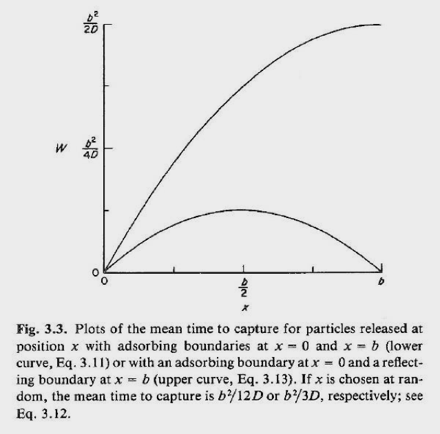
Figure 1. Diagram reproduced from Berg (1993) shows the symmetrical flow behavior of diffusing molecules. It illustrates how displacements, which may be assumed by biologists to be predetermined by design, are impossible due to the overriding effect of random motion driven by collisions of energy, kT.
Forces in the Medium
The scientific literature on the state of water at interfaces is truly vast, and as readers of this journal are aware, expands day-by-day. Yet we must remain mindful of the fact that early contributions to gel research originated from non-biological systems, predating even Frey-Wyssling’s observations. As a student in the 1960s, I learnt of results in the field of soil science obtained already in the 1930s. These reports indicated an extensive ordering effect on the molecules of water in contact with the silica surfaces of hydrophilic clays. For instance, it was already known that montmorillonite and bentonite swell against high imposed pressures (Langmuir, 1938; Norrish, 1954). Hydrophilic vermiculite was shown to produce a crystalline gel at a water to clay ratio of 30 to 1, in which 1 nm thick planar clay wafers are in parallel alignment at an interparticle distance of 50 nm (Walker, 1949, 1960). Already in 1947 Perutz’s group reported that gels of hemoglobin formed “crystals” of 50% water, which appeared to be in ordered layers (Boyse-Watson et al., 1947). Thirteen years later, the term “crystal” was used with confidence when the full X-ray structures of myoglobin and hemoglobin were published (Perutz et al.,1960). For a comprehensive review from the time of those early results see Henniker (1949).
Over the past 60 years, observations of this effect stemming from both biological and non-biological fields have been continually amassed. I have covered this long history elsewhere (Web ref.2). Water that is influenced by the presence of interfaces has been given a variety of names: surface, associated, hydration, vicinal, structured, confined, exclusion-zone. This confused picture stems from the fact that no clear, convincing explanation of its origin has been presented, in turn reflecting the lack of a definitive technique to have emerged from the many fields in which it has been seen, and various methods used to detect it. Israelachvilli’s text “Intermolecular and Surface Forces” (1992) gives a coverage of measurements on clay through to DNA systems gathered already prior to 1990. This text is mathematically oriented; for a non-mathematical alternative consult the major review by Rand and Parsegian (1992). Israelachvilli uses the term “hydration force,” and today it is recognized to exist at all hydrophilic surfaces. I attended the meeting “Biophysics of Water” in Cambridge in 1980 (Franks F and Mathias SF, eds 1981. Wiley Ltd), at which he detailed the apparatus that made the first direct measurement of the force exerted by ordered layers of water between two mica surfaces. Those experiments registered a force normal to the surfaces of up to 1,000 atmospheres. Today the phenomenon is known to be so wide-spread that the physical/chemical nature of the surface does not seem to play a critical role in generating it. Heterogeneous biological tissue and even metals build solute-excluding zones of the pure solvent (Zheng and Pollack, 2003). Research of decades ago (Deryagin, 1966) demonstrated the powerful influence of the silica surface, whereby for example, tightly held water of up to 600 layers were readily formed (Pashley and Kitchener, 1979), which have been shown to explain thermal anomalies in physicochemical properties (Drost-Hansen, 1978). Fast-forwarding now to more recent times, the presence of surfaces and solutes have been shown to have a long-range influence on the H-bond network, which Roke and coworkers call “orientational water” (Chen et al, 2016). Other dielectric response studies show strong anisotropy in confined water as revealed by an order-of-magnitude drop in orientational fluctuations extending as far as 100 nm from silica interfaces (De Luca et al., 2016). The regular array of such layers eases lateral, but restricts normal, surface movement (Dhopatkar et al., 2016). Studies of the long list of unexpected properties has prompted Pollack to theorize that it represents a new physical phase of pure water (2013).
The powerful hydration force was observed also between lipid bilayers as early as the 1970s. As with clay systems, pressures of up to 1,000 atmospheres are needed to force water out of the interlamellar space, or viewed in reverse, to prevent the layered water molecules from pulling in additional water. These macro forces cannot be generated by the chaos of free independent molecular motions generated by kT. If they could, the medium would be readily removed by imposing pressure perpendicular to the layers. As a result, there would be no local forces remaining to orientate proteins embedded in membrane environments, which is needed to direct their highly vectorial functions. A text book example here is the mechanism of ATP synthesis (for an easy concise read, see Boyer, 1999). This multicomponent complex rotates about an axis perpendicular to the membrane plane, whereby each full turn delivers precisely three molecules of ATP. It is driven by the transmembrane displacement of four H+ per ATP. This non-random displacement of H+ supplying the energy for synthesis is an experimental fact known since Mitchell made the discovery of the osmotic drive in 1961. It is not driven by erratic pulses of energy kT on H+ ions, propelling them by chance in the right direction through the membrane.
In addition to Berg’s text cited above, I have also checked other texts recommended for biologists written by prominent physicists (Benedek and Villars, 1974; Nelson, 2008). I found the proposed mechanisms to explain the properties of membrane function quite amazing. For example, one claims that the chaotic influence of thermal motion, kT, on solutes, is “rectified” by membranes, even though as Meister insists, there is no basis in thermodynamics for corrections to molecular motion! To explain the build-up of osmotic forces across membranes, a rectifying force-field is assumed to radiate out of the membrane (Kramer and Myers, 2012). This field operates selectively on molecules of solutes; not water. Thus such claims dismiss decades-long physical, chemical and biochemical research, which the reader can find thoroughly reviewed by Ling (2007). This wide-ranging body of data has clearly established the existence of the hydration force generated by the influence of surfaces on molecules of water; not solutes.
Biological membranes exist in, or better expressed, create a highly anisotropic environment; a space experiencing orthogonal forces controlling the orientation of protein complexes embedded therein. Osada and Gong (1998) offer the thought-provoking suggestion that, because of its solid-like characteristics, the layer of water itself may detect mechanical variations, making it the first link in the response chain. It is a requirement for their function, that membrane proteins be sensitive to small vectorial changes. Just like any agent expecting a stimulus, they must be poised ready for action. This requirement is provided by the geometric arrangement of forces, which prevents randomization. It is therefore expected that nanoscale structural changes due to osmotic, mechanical, electrical and magnetic variations in the surroundings must be able to be detected and amplified, so that resulting displacements occur in the correct direction, and not in any uncontrolled direction resulting from random re-orientations caused by kT. Just like our man-made machines, biological machinery functions with certainty, not chance, so that reliable one-way action is ensured. Clearly, only a structure-based model of the medium can explain how the work we observe reaching up to the cellular level results from the merging of mechanisms down at the molecular level (Watterson, Web ref. 3). Quantum electrodynamic studies have shown co-operative phenomena help to form coherent domains in the structure of liquid water, which are able to mediate successful intermolecular energy transfers by avoiding the disruptive effects of kT (Del Giudice and Giuliani, Web ref. 4).
History of Cytoplasmic Machines
In 1999 the computer corporate giant, IBM, launched the “Blue Gene Project” in a blaze of publicity. Its aim was to solve the “grand challenge problem” (their words) of our time: protein structure. Since their advanced machine, “Blue,” had just defeated the world chess champion, Kasparov, a speedy result was implied. However, in my last correspondence with the project leaders, Ajay Royyuru (Computational Biology Center) and Ruhong Zhou (Research Manager, Protein Science), I understand there has been no progress. We are no closer to understanding the mystery that lies behind protein structure than we were two decades ago.
For readers unfamiliar with the problem, I feel some clarification is needed at this point. I am not referring to solving structure using sequence homology comparison (template based modeling). There are today several algorithms to do that job, which you can carry out on your own PC. Indeed, many volunteers are doing just that. (Interested readers unaware of this cooperative effort, the Community Wide Experiment, or CASP for short, will find it at http://predictioncenter.org). Rather, I refer to elucidating the energetic principles that enable short amino acid sequences of alpha and beta primary structure along the main chain, to fold and pack into a unique stable globule in the 20-30 kDa size range. It is important to emphasize that the stable fold was not predicted by thermodynamicists, and indeed up until the 1990s, it was even considered impossible. Prior to that time, thermodynamics dictated that proteins should adopt the once popular “kicking and screaming stochastic model” (Weber, 1975; Cooper,1976). The image was that the cellular space is full of random thermal motion of solvent and solute molecules, similar to what one would imagine happening to gas molecules in empty space.
In his text “Statistical Mechanics of Chain Molecules,” the Nobel Laurate, P J Flory (1969) presents the theory of polymer structure as understood in the 1970s: the kicking and screaming random coil. Although they are the most important polymers, and the most important solutes on planet Earth, proteins are hardly covered. The suggestion is that their peculiar stability is explained by special internal bonds arising from packing constraints that hold the chain together. When these bonds failed to be found, thermodynamicists invented a new type of bonding called the “hydrophobic bond” (Kauzmann, 1959). And when this special bond failed to materialize, the concept was changed to the “hydrophobic effect” (Tanford, 1968), and soon after to the more scientific sounding “hydrophobic interaction” (Tanford, 1973; Franks, 1975). This term is still in use today and is to be understood as shorthand for the longer expression “an unknown force of special attraction that operates between certain amino acid sequences inside the globule added together with a force of special repulsion that operates between certain amino acid sequences and water outside the globule.”
The approach taken in the Blue Gene Project is the thermodynamically rigorous one of free energy minimization. In simple terms, one steps along the sequence testing the free energy at every twist and turn of each amino acid to arrive at a global minimum. This stochastic approach yields the average fold in the world ruled by kT. At the time of the launch there had been a few hundred X-ray crystal structures published. Today, on the Protein Data Bank there are many tens of thousands available. This means that researchers have at their disposal the precise co-ordinates of millions of atoms all defying kT.
The reason for the failure is that the model ignores the influence of the large-scale ordering occurring in the surroundings. In other words, folding and crystallization emerge out of cooperative phenomena reaching up to the macro level. The cooperativity is already in action down deeper, as shown by the basic gel-sol transition in water. Work in the field of quantum electrodynamics mentioned above has also shown how coherent interactions between water molecules underlie the emergence of collective behavior (Bischof and Del Giudice, Web ref. 5). Confidence in the existence of high-level structuring has seen techniques evolve beyond classical X-ray crystallization. Frey-Wyssling needed the aligned cellulose fiber to see order, but today various computer-assisted analyses reveal as-yet-unexplained energetic changes driving the function of large multimolecular complexes. So now with his words ringing in our ears – there is structure in there – we are confronted by the question of how does biology avoid kT?
Conclusion
The world of biology poses certain basic problems for physics that have remained unrecognized for five decades. These unsolved questions concern the energetics of the cytoplasm at the molecular level. We urgently need input from impartial thermodynamicists to help solve them. Here are three:
1. What do water molecules do when they form a gel?
2. How do they produce the hydration force?
3. What is the explanation of protein structure?
Still today, thermodynamics dictates that proteins do not fold, do not crystallize, and according to Meister do not adopt a definite orientation. Yet each of these claims is contrary to long-standing established experimental facts. Some 20 years ago, I reviewed results from a broad range of fields relevant to these questions (Watterson, 1997), but our knowledge of the state of biological matter at that time has since become overshadowed by the vast amount of new supporting data. I am convinced that continued work on responsive proteins, such as the magnetic biosensors disputed by Meister, will usher in a new era of science, whereby they will be used as tools for probing in-situ the physical properties of biological matter.
References
Anikeeva P, Jasanoff A (2016) Problems on the back of an envelope. eLife 5:e19569.
Benedek GB, Villars FMH (1974) Statistical physics. Vol2. Physics with Illustrative Examples from Medicine and Biology. Addison-Wesley Sec. 2.6.
Berg HC (1993) Random walks in biology. Princeton University Press. p 64.
Boyer PD (1999) What makes ATP synthase spin? Nature 402:247-249.
Boyse-Watson J, Davidson E, Perutz MF (1947) X-ray diffraction pattern from methemoglobin suspensions. Proceedings of Royal Society, London A 191:83-132.
Buxbaum RE, Dennerl T,Weiss S, Heidermann SR (1987) F-actin and microtubule suspensions as indeterminate fluids. Science 235:1511-1514.
Case LB, Waterman CM (2015) Integration of actin dynamics and cell adhesion by a three-dimensional, mechanosensitive molecular clutch. Nature Cell Biology Adv Online Publ: 1-8.
Chen Y, Okur HI, Gomopoulos N, Macias-Romero C, Cremer PS, Petersen PB, Tocci G, Wilkins DM, Liang C, Ceriotti M, Roke S (2016.) Electrolytes induce long-range orientational order and free energy changes in the H-bond network of bulk water. Science Advances 2:e1501891.
Cooper A (1976) Thermodynamic fluctuations in proteins. Proceedings of the National Acadamy of Sciences, USA. 73:2714-2741.
De-Luca S, Kannam SK, Todd BD, Frascoli F, Hansen JS, Daivis PJ (2016) Effect of confinement on the dielectric response of water extends up to mesoscale dimensions. Langmuir 32: 4765-4773.
Deryagin BV (1966) Effect of lyophilic surfaces on properties of liquid films. Discussions of the Faraday Society 42:109-192.
Dhopatkar N, Defante AP, Dhinojwala A (2016) Ice-like water supports hydration forces and eases sliding friction. Science Advances 2:e1600763
Drost-Hansen W (1978) Water at biological interfaces – structural and functional aspects. Phys Chem Liquids 7:243-248.
Feynman RP (1990) QED. Penguin Books, London ch 3.
Flory PJ (1969) Statistical Mechanics of Chain Molecules. Interscience Publishers, p 300-306.
Franks F (1975) The hydrophobic interaction. in Water. A Comprehensive treatise (Plenum, NY) 4:1-64.
Frey-Wyssling A (1940) The submicroscopic structure of the cytoplasm. Journal of Royal Microscopical Society 60:128-139.
Frey-Wyssling A (1948) Submicroscopic morphology of protoplasm. Elsivier, NY.
Gell-Mann M (1994) The Quark and the Jaguar. Little and Brown, London, ch 1.
Gong JP (2006) Friction and librication of hydrogels – its richness and complexity. Soft Matter 2:544-552.
Grasso R, Musumeci F, Gulino M, Scordino A (2018) Exploring the behaviour of water in glycerol solutions using delayed luminescence. PloS One 13:e0191861.
Henniker JC (1949) The depth of the surface zone of a liquid. Reviews of Modern Physics 21:322-341.
Israelachvilli J (1992) Intermolecular and Surface Forces. Academic Press.
Iwamoto H, Inoue K, Yagi N (2006) Evolution of long range myofibrillar crystallinity in insect flight muscle. Proceedings of Royal Society B 273:667-685.
Kauzmann W (1959) Some factors in the interpretation of protein denaturation. Advances in Protein Chemistry 14:1-63.
Kramer EM, Myers DR (2012) Five popular misconceptions about osmosis. Am J Phys 80:694-699.
Langmuir I (1938) The role of attricative and repulsive forces in the formation of tactoids, thixotropic gels, protein crystals and coacervates. Journal of Chemical Physics 6:873-896.
Ling G (2007) Nanoprotoplasm: the ultimate unit of life. Physiol Chem Phys Med NMR 39:111-234.
Meister M (2016) Physical limits to magnetogenetics. eLife 5:e17210.
Nelson P (2008) Biological Physics. WH Freeman and Co. NY. p 258.
Norrish K (1954) The swelling of montmorillonite. Discussions of the Faraday Society 18:120-134.
Osada Y, Gong JP (1998) Soft and wet materials – polymer gels. Advanced Materials 10:827-835.
Palmer KJ, Merrill RC, Ballantyne M (1948) Intercrystalline water in pectinic and pectin acid gels. Journal of American Chemical Society: 570-577.
Pashley RM, Kitchener JA (1979) Mulitlayer adsorption of water on silica. Journal of Colloid and Interface Science 71:491-500.
Perutz MF, Rossmann MG, Cullis AF, Muirhead H, Will G, North ACT (1960) Structure of hemoglobin. Nature 185:416-422.
Pollack GH (2001) Cells, Gels and the Engines of Life. Ebner and Sons, Seattle.
Pollack GH (2013) The Fourth Phase of Water. Ebner and Sons, Seattle. ch 4.
Qin S, Yin H, Yang C, Dou Y, Liu Z, Zhang P, Yu H, Huang Y, Feng J, Hao J, Hao J, Deng L, Yan X, Dong X, ZhaoZ, Jiang T, Wang HW, Luo SJ, Xie C (2016) A magnetic protein biocompass. Nature Materials 15:217–226.
Rand RP, Parsegian VA (1992) The forces between interacting bilayer membranes and the hydration of phospholipic assemblies. in The Structure of Biological Membranes (Yeagle P ed) CRC Press:251-306.
Stanley SA, Kelly L, Latcha KN, Schmidt SF, Yu X, Nectow AR, Sauer J, Dyke JP, Dordick JS, Friedman JM (2016) Bidirectional electromagnetic control of the hypothalamus regulates feeding and metabolism. Nature 531:647-650.
Tanford C (1968) Protein denaturaton. Advances in Protein Chemistry 23:121-282.
Tanford C (1973) The hydrophobic interaction. Wiley, NY ch 1.
Varki A (2017) Biological Roles of Glycans. Glycobiology 27:3-49.
Walker GF (1949) Water layers in vermiculite. Nature 163:726-727.
Walker GF (1960) Hydration of parallel silicate layers. Nature 187:312-314.
Watterson JG (1997) The pressure pixel – unit of life? BioSystems 41:141-152.
Weber G (1975) Energetics of ligand binding to proteins. Advances in Protein Chemistry 29:1-83.
Wheeler MA, Smith CJ, Ottolini M, Barker BS, Purohit AM, GrippoRM, Gaykema RP, Spano AJ, Beenhakker MP, KucenasS, Patel MK, Deppmann CD, Guler AD (2016) Genetically targeted magnetic control of the nervous system. Nature Neuroscience 19:756-761.
Worthington CR (1961) X-ray diffraction studies on large scale molecular structure of insect muscles. Journal of Molecular Biology 3:618-633.
Zheng J-M, Pollack GH (2003) Long-range forces extending from polymer-gel surfaces. Physical Review E68 031408:1-7.
Web References
1. Protein Data Bank, www.pdb101.rcsb.org/browse/molecular-motors.
2. Watterson JG (2017) Comment on Meister’s Article “Physical Limits to Magnetogenetics” https://elifesciences.org/profiles/9axyx4gy
3. Watterson JG www.thewaterpixel.com;
www.lifeenergyscience.it/journal.htm
4. Del Giudice E, Giuliani L (2010) Coherence in water and the kT problem in living matter http://www.icems.eu/papers/ramazzini_library5_part1.pdf
5. Bischof M, Del Giudice E (2013) Communication and the emergence of collective behavior in living systems. http://dx.doi.org/10.1155/2013/987549